
Image Processing and Cancer Detection
Histopathological Sections
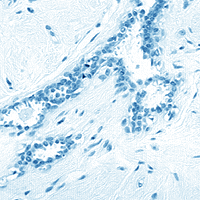
Malignant lesions are readily distinguishable from normal tissue samples.
|
|
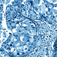
Under high magnification, changes become apparent in the nuclei of the cancerous tissue. As lesions develop, uncondensed chromatin becomes more pronounced, appearing darker than normal tissue. These densely clustered granules allow less light to pass through them when trans-illuminated.


in cancer cells (R), nuclear chromatin patterns vary from the norm
Images of nuclei are isolated from the tissue background during a process known as image segmentation. Digital analysis of nuclear images is the key to unlocking information hidden to the unaided eye.

|

|

![]()


The University of Arizona
March 26, 1998
denicew@u.arizona.edu
http://www.biology.arizona.edu
All contents copyright © 1998. All rights reserved.