Image Processing and Cancer Detection
Classification of Nuclei
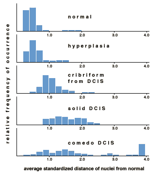
The final phase in this process of quantitative analysis is to incorporate all measurable features of a sample into a single cell profile. Each of the 99 features is analyzed according to its standardized distance from a corresponding normal value. The "Average Standardized Distance from Normal," then, is an average of each of these 99 values, where distance is measured in standard deviations.
Once a profile has been generated, lesions can be classified according to degree of abnormality and grade of progression. Highly specific determinations can be made at this point regarding optimum patient management.

Although a pathologist would diagnose both of these lesions as cancerous, only through the processes of digital and statistical analysis can this marked difference in severity be illustrated.
Whereas traditional cancer survival statistics are based primarily on life span averages for patients with similar lesions, application of image processing technology could make available a lesion-specific prediction that is more accurate than has ever before been possible.
The University of Arizona
March 26, 1998
denicew@u.arizona.edu
http://www.biology.arizona.edu
All contents copyright © 1998. All rights reserved.


